BMRB Entry 11026
Click here to enlarge.
PDB ID:
Entry in NMR Restraints Grid
Validation report in NRG-CING
Chem Shift validation: AVS_full, LACS, SPARTA
BMRB Entry DOI: doi:10.13018/BMR11026
MolProbity Validation Chart
NMR-STAR file interactive viewer.
NMR-STAR v3 text file.
NMR-STAR v2.1 text file (deprecated)
XML gzip file.
RDF gzip file.
All files associated with the entry
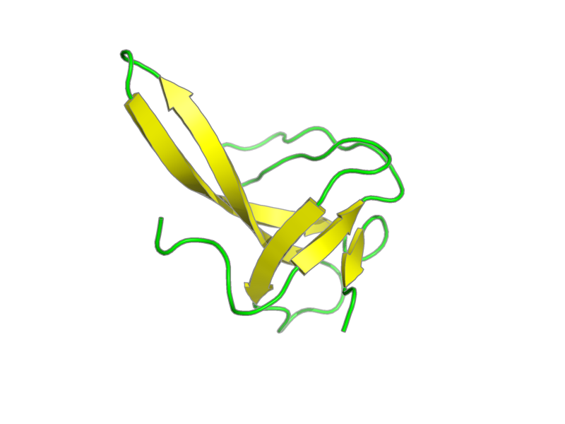
Title: SOLUTION STRUCTURE OF ALPHA-SPECTRIN SH3-BERGERAC FROM CHICKEN PubMed: 11478866
Deposition date: 2008-01-17 Original release date: 2009-05-21
Authors: Kutyshenko, Victor; Prokhorov, Dmitry; Timchenko, Maria; Kudrevatykh, Yuri; Fedyukina, Daria; Guschina, Lyubov; Khristoforov, Vladimir; Filimonov, Vladimir
Citation: Viguera, A.; Serrano, L.. "Bergerac-SH3: "frustation" induced by stabilizing the folding nucleus." J. Mol. Biol. 311, 357-371 (2001).
Assembly members:
alpha-spectrin SH3-bergerac, polymer, 70 residues, 8068.312 Da.
Natural source: Common Name: Chicken Taxonomy ID: 9031 Superkingdom: Eukaryota Kingdom: Metazoa Genus/species: Gallus gallus
Experimental source: Production method: recombinant technology Host organism: Escherichia coli
Entity Sequences (FASTA):
alpha-spectrin SH3-bergerac: MDETGKELVLALYDYQEKSP
REVTMKKGDILTLLNSTNKD
WWKVEVKATANGKTYERQGF
VPAAYVKKLD
- assigned_chemical_shifts
| Data type | Count |
| 13C chemical shifts | 320 |
| 15N chemical shifts | 69 |
| 1H chemical shifts | 489 |
Additional metadata:
Assembly:
| Entity Assembly ID | Entity Name | Entity ID |
|---|---|---|
| 1 | alpha-spectrin SH3-bergerac | 1 |
Entities:
Entity 1, alpha-spectrin SH3-bergerac 70 residues - 8068.312 Da.
This is the mutant of alpa-spectrin wild type SH3-domain. The difference between this mutant and wild type protein consists of 10 extra residues placed between 45th and 48th polypeptides' residues.
| 1 | MET | ASP | GLU | THR | GLY | LYS | GLU | LEU | VAL | LEU | |
| 2 | ALA | LEU | TYR | ASP | TYR | GLN | GLU | LYS | SER | PRO | |
| 3 | ARG | GLU | VAL | THR | MET | LYS | LYS | GLY | ASP | ILE | |
| 4 | LEU | THR | LEU | LEU | ASN | SER | THR | ASN | LYS | ASP | |
| 5 | TRP | TRP | LYS | VAL | GLU | VAL | LYS | ALA | THR | ALA | |
| 6 | ASN | GLY | LYS | THR | TYR | GLU | ARG | GLN | GLY | PHE | |
| 7 | VAL | PRO | ALA | ALA | TYR | VAL | LYS | LYS | LEU | ASP |
Samples:
sample_1: alpha-spectrin SH3-bergerac, [U-98% 13C; U-98% 15N], 2.5 mM; sodium acetate, [U-99% 2H], 25 mM; sodium azide 0.03%; H2O 90%; D2O, [U-99% 2H], 10%
sample_conditions_1: ionic strength: 25 mM; pH: 4; pressure: 744 mmHg; temperature: 298 K
Experiments:
| Name | Sample | Sample state | Sample conditions |
|---|---|---|---|
| 2D 1H-15N HSQC | sample_1 | isotropic | sample_conditions_1 |
| 3D HNCACB | sample_1 | isotropic | sample_conditions_1 |
| 3D C(CO)NH | sample_1 | isotropic | sample_conditions_1 |
| 3D HCCH-TOCSY-ali | sample_1 | isotropic | sample_conditions_1 |
| 3D HCCH-TOCSY-aro | sample_1 | isotropic | sample_conditions_1 |
| 3D 1H-15N TOCSY | sample_1 | isotropic | sample_conditions_1 |
| 3D 1H-15N NOESY | sample_1 | isotropic | sample_conditions_1 |
| 3D 1H-13C NOESY-ali | sample_1 | isotropic | sample_conditions_1 |
| 3D 1H-13C NOESY-aro | sample_1 | isotropic | sample_conditions_1 |
| 3D HNCO | sample_1 | isotropic | sample_conditions_1 |
Software:
CARA, Rochus Keller and Kurt Wuthrich - chemical shift assignment, data collection, processing
TALOS, Cornilescu, Delaglio and Bax - dihedral angles calculation
PREDITOR, PREDITOR - dihedral angles calculation
CYANA v2.1, Guntert, Mumenthaler and Wuthrich - structure solution
Molmol, Koradi, Billeter and Wuthrich - Data analysis
NMR spectrometers:
- Bruker Avance 600 MHz
Download simulated HSQC data in one of the following formats:
CSV: Backbone
or all simulated shifts
SPARKY: Backbone
or all simulated shifts