BMRB Entry 11102
Click here to enlarge.
PDB ID:
Entry in NMR Restraints Grid
Validation report in NRG-CING
Chem Shift validation: AVS_anomalous, AVS_full, LACS
BMRB Entry DOI: doi:10.13018/BMR11102
MolProbity Validation Chart
NMR-STAR file interactive viewer.
NMR-STAR v3 text file.
NMR-STAR v2.1 text file (deprecated)
XML gzip file.
RDF gzip file.
All files associated with the entry
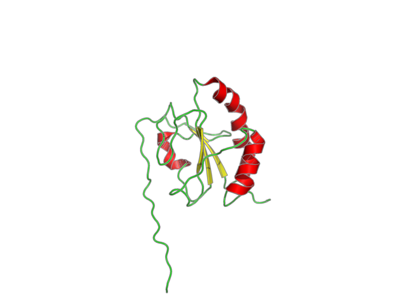
Title: The solution structure of the second thioredoxin domain of human Protein disulfide-isomerase A3
Deposition date: 2010-02-18 Original release date: 2011-02-17
Authors: Tochio, N.; Koshiba, S.; Inoue, M.; Kigawa, T.; Yokoyama, S.
Citation: Tochio, N.; Koshiba, S.; Inoue, M.; Kigawa, T.; Yokoyama, S.. "The solution structure of the second thioredoxin domain of human Protein disulfide-isomerase A3" . ., .-..
Assembly members:
thioredoxin domain, polymer, 142 residues, Formula weight is not available
Natural source: Common Name: human Taxonomy ID: 9606 Superkingdom: Eukaryota Kingdom: Metazoa Genus/species: Homo sapiens
Experimental source: Production method: cell free synthesis Host organism: E. coli - cell free
Entity Sequences (FASTA):
thioredoxin domain: GSSGSSGFDGNLKRYLKSEP
IPESNDGPVKVVVAENFDEI
VNNENKDVLIEFYAPWCGHC
KNLEPKYKELGEKLSKDPNI
VIAKMDATANDVPSPYEVRG
FPTIYFSPANKKLNPKKYEG
GRELSDFISYLQREATSGPS
SG
- assigned_chemical_shifts
| Data type | Count |
| 13C chemical shifts | 611 |
| 15N chemical shifts | 132 |
| 1H chemical shifts | 925 |
Additional metadata:
Assembly:
| Entity Assembly ID | Entity Name | Entity ID |
|---|---|---|
| 1 | thioredoxin domain | 1 |
Entities:
Entity 1, thioredoxin domain 142 residues - Formula weight is not available
| 1 | GLY | SER | SER | GLY | SER | SER | GLY | PHE | ASP | GLY | ||||
| 2 | ASN | LEU | LYS | ARG | TYR | LEU | LYS | SER | GLU | PRO | ||||
| 3 | ILE | PRO | GLU | SER | ASN | ASP | GLY | PRO | VAL | LYS | ||||
| 4 | VAL | VAL | VAL | ALA | GLU | ASN | PHE | ASP | GLU | ILE | ||||
| 5 | VAL | ASN | ASN | GLU | ASN | LYS | ASP | VAL | LEU | ILE | ||||
| 6 | GLU | PHE | TYR | ALA | PRO | TRP | CYS | GLY | HIS | CYS | ||||
| 7 | LYS | ASN | LEU | GLU | PRO | LYS | TYR | LYS | GLU | LEU | ||||
| 8 | GLY | GLU | LYS | LEU | SER | LYS | ASP | PRO | ASN | ILE | ||||
| 9 | VAL | ILE | ALA | LYS | MET | ASP | ALA | THR | ALA | ASN | ||||
| 10 | ASP | VAL | PRO | SER | PRO | TYR | GLU | VAL | ARG | GLY | ||||
| 11 | PHE | PRO | THR | ILE | TYR | PHE | SER | PRO | ALA | ASN | ||||
| 12 | LYS | LYS | LEU | ASN | PRO | LYS | LYS | TYR | GLU | GLY | ||||
| 13 | GLY | ARG | GLU | LEU | SER | ASP | PHE | ILE | SER | TYR | ||||
| 14 | LEU | GLN | ARG | GLU | ALA | THR | SER | GLY | PRO | SER | ||||
| 15 | SER | GLY |
Samples:
sample_1: thioredoxin domain, [U-13C; U-15N], 1.3 mM; d-Tris-HCl 20 mM; NaCl 100 mM; d-DTT 1 mM; NaN3 0.02%; H2O 90%; D2O 10%
condition_1: ionic strength: 120 mM; pH: 7.0; pressure: 1 atm; temperature: 296 K
Experiments:
| Name | Sample | Sample state | Sample conditions |
|---|---|---|---|
| 3D 1H-15N NOESY | sample_1 | isotropic | condition_1 |
| 3D 1H-13C NOESY | sample_1 | isotropic | condition_1 |
Software:
xwinnmr v3.5, Bruker - collection
NMRPipe v20031121, Delaglio, F. - processing
NMRView v5.0.4, Johnson, B.A. - data analysis
Kujira v0.955, Kobayashi, N. - data analysis
CYANA v2.0.17, Guntert, P. - structure solution
NMR spectrometers:
- Bruker AVANCE 900 MHz
Download simulated HSQC data in one of the following formats:
CSV: Backbone
or all simulated shifts
SPARKY: Backbone
or all simulated shifts